Measurement of these biophotons remains challenging due to the need to both remove all stray light, to minimize the noise within the measurement system and to avoid motion artifacts. This degree of optical isolation means that measurements are currently performed in a darkroom. Rather than performing this procedure in darkness, however, the region of interest and biophoton collector can be shrouded, but care must be taken to avoid external light scattering through the tissue and reaching the detector. Thermal noise in the measurement system can be reduced by operating at cryogenic temperatures, either by using liquid nitrogen or Peltier cooling. The patient too must be held at as constant a temperature to avoid a change in the flux of thermal photons. Once a system has been assembled, imaging takes tens of minutes due to the tiny photon fluxes available, currently requiring the patient to remain absolutely still throughout to avoid motion artifacts that would dominate over the signal from cancerous cells.
None of these challenges is insurmountable. This could be a real step-change for cancer diagnostics as low-light measurement is continually improving. If measurement is performed in-vivo, for example by using an endoscope, ambient light levels would be substantially reduced relaxing this constraint significantly. Integration times are unlikely to be reduced to less than a few minutes, but instead, patient motion could be mitigated through secure mounting of the detector onto the patient during image acquisition and algorithmic adaptations to correct for motion in the image.
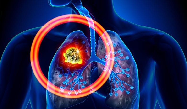
After ten years of academic development, measurement of spontaneous ultraweak photons from cancerous cells is now at the point where the fundamental science is sufficiently well-established to allow the development of a commercial cancer imaging device without the need for labeling such as using a fluorescent dye and is thus able to provide a really direct measurement.